
Photo by: Tom Robertson Photography
The topic of bone stress injuries (BSIs) is very complex, and research continues to investigate these injuries so they can be better prevented and appropriately managed. BSIs are quite common among athletes, comprising over 10% of all sport-related injuries, account for up to 20% of all injuries treated in sports medicine clinics, and account for up to 30% of all running related injuries (1-3). I know too many stories of runners who have unsuccessfully tried to run through bone stress injuries, either because they held off too long on seeking diagnosis and treatment, or because their healthcare provider misdiagnosed the injury. The goal of this discussion is to provide a summary of the risk factors, classic symptoms, common locations, and importance of appropriate diagnosis of BSIs. Readers should come away with a better understanding of when they might need to seek help from a qualified medical professional so they may return to running as soon as possible.
What is a bone stress injury?
Simply put, a BSI occurs when a bone is unable to withstand the repetitive loading that is being applied to it. Although they are often referred to as “stress fractures,” these injuries occur along a pathology continuum ranging from a “stress reaction,” involving inflammation of the periosteum and bone marrow, to actual stress fracture where distinct fracture lines are present (2).
Why do bone stress injuries occur?
Bone physiology is extremely complex, but it is important to understand the basics in order to fully respect why a BSI may occur and how to address the underlying cause. Although we think of bones as solid static structures, on a microscopic level, your body is continually breaking down and re-building your bones. In addition to providing structural support for your body, bones are an important calcium storage system. Calcium is essential in many vital bodily functions such as muscle contraction, blood clotting, enzyme function, and heart rhythm. Your body is continually borrowing from the bone calcium stores by breaking down the bone (resorption), and then rebuilding it. In a healthy individual, bone rebuilding occurs at approximately the same, or even higher rate than it is being broken down. This is called skeletal homeostasis and occurs in all individuals, even those who are not exercising. Bone will also remodel specifically in response to being loaded. When a bone experiences stress via either external loads (impact of your body contacting the ground) and/or internal loads (the force applied to the bone by the contraction of the muscles that attach to it), load-induced microtrauma occurs. When the body systems are functioning appropriately, the additional loading is good for stimulating bone growth and remodeling, resulting in a bone that is rebuilt stronger and more acclimated to higher loads.
The following conditions must be in place in order for healthy bone remodeling to occur:
Adequate energy availability: To put it very simply, this means eating enough calories to support your workouts AND your vital body functions. Sometimes runners don’t realize just how much they need to eat in order to support their activity level. Sometimes, runners purposefully restrict food intake with hopes of losing weight in order to to look a certain way, or with the hopes that they will perform better. When compared to athletes who are not training in a state of low energy availability, physically active individuals in a state of low energy availability tend to eventually present with lower bone mass, altered bone metabolism (favoring bone resorption), reduced bone strength and increased risk for BSIs. Being in a state of low energy availability for even 5 days appears to negatively affect the balance of bone resorption and bone formation (4). Low energy availability seems to be the root cause for many physiological and performance dysfunctions including several of the points discussed below. It is a very large and important topic worthy of ten of it’s own blog posts. If you would like to read more on relative energy deficiency in sport (RED-S), I have provided some links to good resources below:
Lecture by Dr. Kathryn Ackerman on the Triad and RED-S that provides a great summary of the current literature on the subject. It specifically addresses bone health beginning at 3:45.
Adequate hormone circulation: Low energy availability is also correlated with a decrease in several hormones that are essential for bone health including the “sex hormones”(estrogen and testosterone), and has a negative effect on some metabolic hormones such as leptin, IGF-1, and peptide YY (5,6). Many of these hormonal changes likely occur to conserve energy for more important bodily functions or to use the body’s energy reserves for vital processes (7).
Regular menstruation (in females): Females who stop having regular menstrual cycles (amenorrhea) or experience < 9 menstrual cycles in a year (oligomenorrhea) have demonstrated decreased BMD, altered bone microarchitecture and bone turnover markers, decreased estimates of bone strength and increased risk for bone stress injuries compared with eumenorrhoeic athletes (6,7). This is likely due to a combination of the reduced hormone circulation that results in amenorrhea as well as state of low energy availability that often occurs simultaneously(5). The interrelationship of decreased energy availability, menstrual dysfunction, and poor bone health, is known as the female athlete triad. In women who participate in sports that emphasize aesthetics or leanness, such as ballet or running, the prevalence of amenorrhea can be as high as 69%, compared with 2% to 5% in the general population (8).
Males don’t menstruate, but…: Although males do not experience menstrual cycles, they can experience a similar condition as the female athlete triad called exercise-related male hypogonadism, which is characterized by a deficiency in the production of testosterone – a critical male reproductive hormone. Like the Triad, it is often correlated with a state of low energy availability, and decreased bone mineral density. Symptoms are less obvious in males, and blood-work is necessary for proper diagnosis, but one of the more noticeable symptoms is loss of morning erections and/or decreased libito (9,10). See link below for a great resource to better understand hypothalamic amenorrhea and male hypogonadism:
No Period, Now What: Website with may blog posts on this topic. You may also purchase the book here as well.
Hypogonadism in Exercising Males: Dysfunction or Adaptive-Regulatory Adjustment? Hackney 2020
Adequate vitamin D and calcium: Even when a runner is taking in enough calories to support training loads, adequate levels of vitamin D and calcium are essential for good bone health. Low levels result in decreased bone strength and higher risk for BSI (5).
Appropriate training load progression: Too much load applied to a bone that is not accustomed to being loaded (i.e. increasing running mileage too quickly) will increase risk of BSI (3). Bones are dynamic and will remodel over time to acclimate to progressively higher training loads, but too much too soon will result in eventual injury. Just like a chair that is build to withstand 200 lbs of weight may break if 300 lbs of weight is placed on it. Training load management is also a very complex topic and important for prevention and treatment of all running related injuries. Below are links to a few articles I have written on these topics:
Avoid Training Error, Avoid Injury: A post I wrote on the Endurance Physio Blog in 2018.
Injury Prevention Though Training Load Management: An article I wrote for The Cycling House in 2019. This one is more focused on cyclists and triathletes, but the concepts are very applicable to runners.
Adequate rest: It takes time to repair the bone tissue. Training will provide a stimulus for bone remodeling to occur, but too many successive training bouts without recovery time will not allow for the rebuilding phase to occur and the bone will break down at a faster rate than it can be rebuilt.
Adequate sleep: Sleep deprivation is associated with a higher risk for reduced bone health due to alterations in the balance between bone resorption and remodeling (11).
Where do BSIs usually occur?
The most common sites for BSI in runners include the medial tibia, femoral shaft, fibula, calcaneus, and metatarsal bones, but can also occur in the pelvis, sacrum, lumbar spine, femoral neck, anterior tibia, and navicular (see images below for an idea of some of these common anatomical locations where pain may be experienced) (2,3). Any bone could technically become injured, but these are the ones that are most likely to occur.
Low risk vs high risk locations
BSIs are categorized as being in either a “low risk” or “high risk” site depending on the location. “High risk” sites tend to be more susceptible to fracture propagation with more severe consequences if fracture does complete, and they are more susceptible to delayed union. These require a higher level of attention and may require surgical intervention. “Low risk” sites have more reliable healing patterns and low risk for fracture completion. The sites for high risk fractures include the fifth metatarsal, the anterior tibia, the navicular, the femoral neck, the patella, the medial malleolus, the talar neck, and the first metatarsal sesamoids (2,3,12).

Warden et al. J Orthop Sports Phys Ther. 2014
Where would it hurt? (not all locations are shown below)

Medial tibia: usually in the distal 1/3 and will be particularly painful in one small area as opposed to along the entire tibial shaft

Navicular (High risk due to poor blood supply and non-union potential)
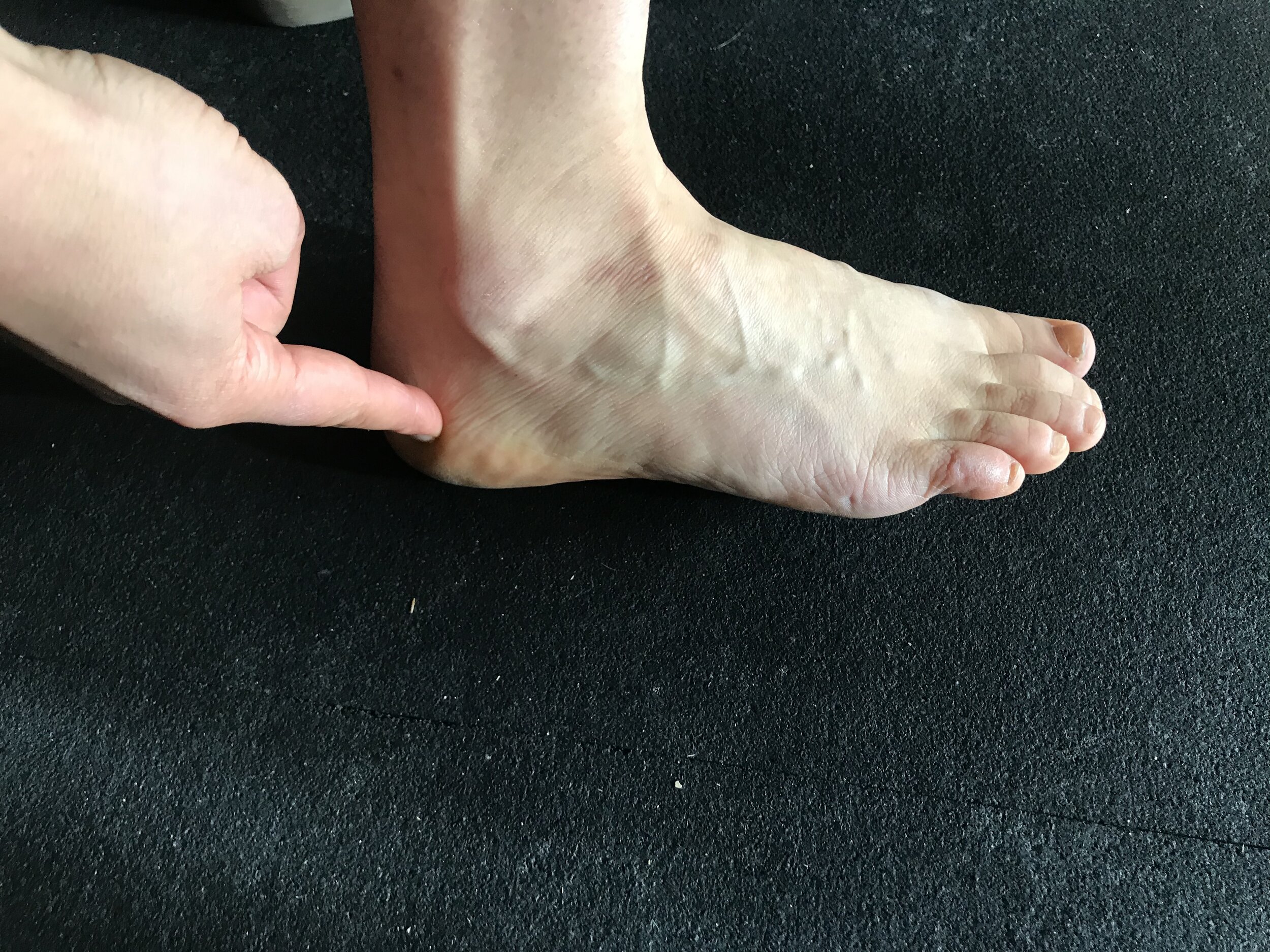
Calcaneus (Some sources categorize as high risk)

Fifth metatarsal (High risk due to poor blood supply and non-union potential)

Sacrum or spine. Not high risk, but should raise suspicion for potential of low energy availability or poor bone health

Fibula

2nd metatarsal

Femoral shaft
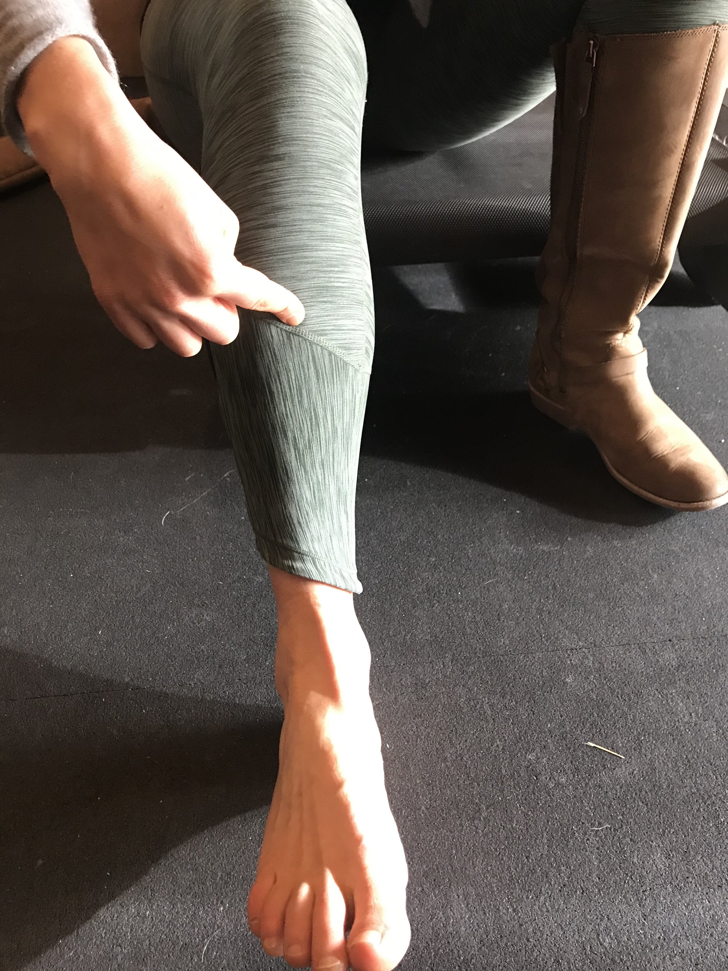
Anterior tibia (High risk due to vulnerability to fully fracture and associated complications if it does)
Not pictured: Femoral neck, and pubic ramus which present as deep groin pain. (Just couldn’t bring myself to publish a crotch-shot :)
(High risk due to vulnerability to fully fracture and associated complications if it does)
How are BSIs diagnosed?
BSIs are diagnosed based on symptom quality, location, and diagnostic imaging. Common symptoms include:
Pain reproduction with palpation to the injured bone: This is difficult if the bone is not superficial, such as femoral neck.)
Pain at rest or at night: in more severe cases
Palpable bone callus and/or visible swelling: Bone callus may be present after the BSI has been present for several weeks. The callus formation is part of the bone healing process.
Symptom reproduction with impact and/or forceful activation of the muscles that attach to the bone
Pain that increases throughout a duration of a run: Often there are tendons that run in the vicinity of the suspect bone, making it difficult to differentiate tendon from bone. But unlike tendon injuries, bone pain tends to gradually increase throughout the duration of a run whereas tendon pain generally decreases once it is warmed up.
Pain that results in limping or inability to tolerate running at all. Bone pain may initially be subtle enough that running remains tolerable without gait alterations, but generally will increase in severity to the point where running is intolerable.
If a BSI is suspected, recommendation is complete avoidance of painful activity until the bone is fully healed, as it will not heal if it continues to be loaded. This is different from tendon injuries, where some pain during running is often allowed, as tendon responds well to loading compared to complete rest. This will be further discussed in part two of this blog series.
When is diagnostic imaging indicated?
If a BSI is suspected, the following algorithm concept may be used in order to guide if, when, and what type of imaging is necessary (13):

Wright et al. 2015
Imaging is often an part of the diagnostics required for appropriate treatment of BSIs and will allow for a more accurate diagnosis and prognosis, and is paramount if the suspected BSI is in a high risk location. X-rays will often come up negative unless the BSI is quite severe and has been present for several weeks. MRI is the most sensitive and specific way to identify the presence and severity of a BSI. One way to help classify the severity and prognosis of the injury is use of the Fredericson classification system, which is based off or MRI results (14):
Grade 1: Periosteal edema only
Grade 2: Bone marrow edema (visible only on T2 weighted sequences)
Grade 3: Bone marrow edema (visible on T1 and T2 weighted sequences)
Grade 4: (4a) Multiple discrete areas of intracortical signal changes; (4b) Linear areas of intracortical signal change correlating with a frank stress fracture
It is important to understand that an X-ray will not detect the lower grade bone stress injuries. Understanding the severity/grade of BSI is important for helping your medical provider devise an optimal treatment plan, and for the injured athlete to gain a better understanding of how long it may take to return to running. Approximate return to sport timelines for male and female track and cross-country runners have been reported as follows (14):
Grade 1: 11.4 weeks (+/- 4.5 weeks)
Grade 2: 13.5 weeks (+/- 2.1 weeks)
Grade 3: 18.8 weeks (+/- 2.9 weeks)
Grade 4: 31.7 weeks (+/- 3.7 weeks)
You can see that BSIs often result in a long time away from sport, so early detection is key for expediting this relatively long recovery, especially if the suspect region is in one of the more “high risk” sites that was discussed earlier. Not only is appropriate diagnosis essential for a proper rehabilitation to occur, it is important for individuals to know if they have had a BSI, since they can be a signal of decreased bone health, underlying energy deficiency, or endocrine dysfunction. Multiple BSIs and/or BSIs that occur in certain regions such as the femoral neck, pelvis, sacrum, spine (due to the type of bone present in these regions) should be a reason for receiving a bone density scan and blood work (15). Make sure to consult with a sports medicine doctor who can help decide when further testing is needed to minimize risk for future BSIs.
Click here for part two, which will discuss management, rehabilitation, and return to running following bone stress injury
Please feel free to comment or contact me at endurancephysioanya@gmail.com with any questions. Thanks for reading!
References
Fredericson M, Jennings F, Beaulieu C, and Matheson G. Stress fractures in athletes. Top Magn Reson Imaging. 2006; 17(5):309-25.
Song SH, Koo JH. Bone Stress Injuries in Runners: a Review for Raising Interest in Stress Fractures in Korea. J Korean Med Sci. 2020;35(8):e38.
Warden SJ, Davis IS, Fredericson M. Management and prevention of bone stress injuries in long-distance runners. J Orthop Sports Phys Ther. 2014;44(10):749–765.
Papageorgiou M, Dolan E, Elliott-Sale KJ, Sale C. Reduced energy availability: implications for bone health in physically active populations. Eur J Nutr. 2018;57(3):847–859.
Goolsby MA, Boniquit N. Bone Health in Athletes. Sports Health. 2017;9(2):108–117.
Ackerman KE, Cano Sokoloff N, DE Nardo Maffazioli G, Clarke HM, Lee H, Misra M. Fractures in Relation to Menstrual Status and Bone Parameters in Young Athletes. Med Sci Sports Exerc. 2015;47(8):1577–1586.
Mountjoy M, Sundgot-Borgen JK, Burke LM, et al. IOC consensus statement on relative energy deficiency in sport (RED-S): 2018 update. Br J sports med. 2018;52:687-697.
Nazem TG, Ackerman KE. The female athlete triad. Sports Health. 2012;4(4):302–311.
Tenforde AS, Barrack MT, Nattiv A, et al. Parallels with the female athlete triad in male athletes. Sports Med. 2016;46:171–82.
Hackney, AC. Hypogonadism in Exercising Males: Dysfunction or Adaptive-Regulatory Adjustment? Front Endocrinol. 2020.
Staab JS, Smith TJ, Wilson M, Montain S, Gaffney-Stomberg, E. Bone turnover is altered during 72 h of sleep restriction: a controlled laboratory study. Endocrine. 2019;65: 192–199.
Chen YT, Tenforde AS, Fredericson M. Update on stress fractures in female athletes: epidemiology, treatment, and prevention. Curr Rev Musculoskelet Med. 2013;6(2):173–181.
Wright AA, Hegedus EJ, Lenchik L, Kuhn KJ, Santiago L, Smoliga JM. Diagnostic Accuracy of Various Imaging Modalities for Suspected Lower Extremity Stress Fractures: A Systematic Review With Evidence-Based Recommendations for Clinical Practice. Am J Sport Med. 2016;44(1):255–263.
Nattive A, Kennedy G, Barrack MT, Abdelkerim A, Goolsby MA, Arends JC, Seeger LL. Correlation of MRI Grading of Bone Stress Injuries with Clinical Risk Factors and Return to Play: A 5-year prospective study in collegiate track and field athletes. Am J Sports Med. 2013; 41(8): 1930-1941.
Marx RG, Saint-Phard D, Callahan LR, Chu J, Hannafin JA. Stress Fracture Sites Related to Underlying Bone Health in Athletic Females. Clin J Sport Med. 2001;11:73-76.